
Aetiologies and Characterisations of Epilepsies
It has been demonstrated, by electro-convulsive therapy, that any human brain is capable of generating a convulsive seizure when external current is applied to the appropriate neural circuits. Epileptic seizures are believed in most instances to stem from an atypical hypersynchronisation of activity between neural paths as a consequence of excessive excitation, lack of inhibition, or structural anomalies within the involved neurons. Thus there are a diverse range of possible neural defects that may contribute toward an increased susceptability to epeleptic seizures.
The manifestations presented by seizures depend on their localisations. The different form of seizures and epileptic syndromes have been well characterised and classifications of each can be found at within the corresponding pages of this website.
The Neuron as the cause of excess activity
The bellow are mediated by proteins present within the neuron, as such, defects contributing to a predisposition to seizures may lie within the proteins themselves, their levels of expression within the cell or at the plasma membrane, their localizations, or their regulation by other proteins and/or molecules. It should also be noted that many effects within the neuron, such as those performed by metabotropic receptors, occur through long signaling pathways involving the interactions of numerous proteins and molecules, each step being a possible candidate for defects resulting in excessive neuronal activity. For the purpose of revision, videos giving a good overview of action potential propagationand synaptic transmission can be found through the following links.
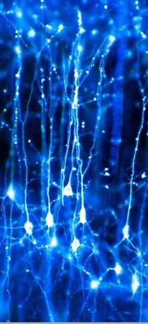 |
| Image of pyramidal neurons by Dr Jonathan Clarke (Wellcome Images images@wellcome.ac.uk) from https://www.flickr.com/photos/lorelei-ranveig/2294885580/ under the creative commons liscence |
The resting membrane potential establishes an important boundary to neural firing, permitting only those stimuli strong enough to bridge the voltage gap between it and the activation threshold to generate an action potential. Ion transporter proteins present in the plasma membrane maintain the required balance the different intracellular ion concentrations to maintain an appropriate resting potential. Some cortical slices from epilepsy sufferers show an inability to accumulate extracellular potassium and reduced sodium – potassium exchange, a consequence of what is likely a defective sodium-potasium ATPase exchanger. This results in a higher than normal resting potential, permitting smaller stimuli to yield neuron firing.
A refractory period between action potentials, created by the voltage dependant inactivation of the sodium channels that mediate the rising phase of an action potential, prevents the summation of activity and limits the frequency of firing. Some models of epilepsy exhibit a reduced refractory period, permitting greater frequencies of neuronal firing.
Ion channels are tightly gated, opening only within defined voltages or on ligand binding, ensuring only appropriate ionic fluxes. Heritable or aquired defects may result in inappropriate ionic fluxes within certain contexts and consequently create windows of opportunity where contributions to spontaneous action potentials can be made. Incorrect regulation of ion channels by other protiens or molecules may create simillar windows. For instance magnesium inhibits NMDA calcium channels in the postsynaptic membrane, thus low levels permit an increase in calcium entry.
Neurotransmitter receptors found on the postsynaptic membrane are essential in correctly mediating the transmission of an action potential from the pre-synaptic neuron to the post. Excitatory neurotransmitter receptors facilitate the entry Ca2+ contributing toward EPSPs, whereas inhibitory neurotransmitter receptors activate chloride channels or potassium channels, resulting in hyperpolarisation of the neuron (negative contribution toward EPSP). The interactions within and between these activated receptors is highly complex, and complications with any can dramatically alter the effects of others. There is evidence for gain of function in post-synaptic excitation and/or a loss of function in post-synaptic inhibition in some forms of epilepsy created by defects in such receptors.
Abnormal levels or patterning of neurtransmitter release too may yield an increased likelyhood of postsynaptic activity.
Long term potentiation of neurons may be an important method of facilitating seizures, by creating a positive feedback loop which lowers the activation threshold for generating action potentials of effected neurons for extended periods of times.
Anatomical Anomalies
Damage to the cerebral hemispheres or abnormal neural development may interfere with normal neural interactions, facilitating atypical neuronal inputs or lack of, possibly creating subsequent novel patterns of activity. Such features may be conducive of an increased susceptibility to epileptic seizures.
Recurring trends in the aetiologies of focal Epelepsies
Mechanisms of inducing excess excitation
- Enhanced activation of NMDA receptors – NMDA receptors are one of the postsynaptic mediators of glutamates action, the major excitatory neurotransmitter of the CNS.
- Increased synchronicity due to novel emphatic interactions – Electrical activity may propagate between neighbouring neurons within laminar structures, such as the hippocampus, by non synaptic means such as gap junctions or electrical field created with synchronous neural activity.
- Reccurent excitatory collaterals which act to increase activity and / or synchronicity.
Mechanisms of decreasing inhibition
- Defective GABA pathways – As the major inhibitory neurotransmitter of the CNS, GABA mediates most inhibitory post synaptic inputs in the brain. Thus congenital defects within any part of its signalling pathway or its regulation will yield wide scale reductions in the brains ability to inhibit neuronal activity. Simillar, though localised, effects are seen with damage to GABAergic and their associated neurons.
Defects inducing cell Death
- Defective calcium signalling – Calcium plays crucial roles as a second messenger in innumerable signalling pathways including those involved in neuronal growth and survival. Experiments have shown the addition of calcium chelating proteins prevents the deterioration of interneurons which characteristically die some epilepsies, suggesting defective intracellular calcium buffering within these.